Anatomy: Small Animal

Welcome to small animal anatomy!
In this section we will be going through some of the major body systems of dogs and cats, such as the cardiovascular system, the musculo-skeletal system, the central nervous system, the digestive system and the respiratory system.
Canine Skeleton

https://zooarch.illinoisstatemuseum.org/content/types-and-parts-bones
Canine Skeleton
The canine skeleton is divided into two regions: THE AXIAL SKELETON and THE APPENDICULAR SKELETON.
The axial skeleton consists of the vertebral column and the rib cage. The appendicular skeleton consists of the pectoral girdle/limb and the pelvic girdle/limb. The vertebral column is a chain comprised of a varying number of vertebrae.
Dogs have 7 cervical vertebrae, 13 thoracic vertebrae, 7 lumbar vertebrae, 3 sacral vertebrae, and 20-23 coccygeal vertebrae.
The vertebral column has 3 functions: protection of the spinal cord, weight bearing and muscle insertion, and movement.


Feline Skeleton

https://quizlet.com/373820684/cat-skeleton-diagram/
Cats and dogs have very similar skeletons because they are both carnivores. So once you know the skeletal anatomy of dogs it is easy to apply that anatomy to cats! Look at the image to see a cat skeleton. Does it look similar to the dog skeleton to you?

Feline Distal Limbs
This image shows the skeletal structure of a cat's distal limb including its carpal bones, metacarpals and phalanges (aka its wrist, hand, and fingers). Cats and dogs both have 5 phalanges or fingers on their front legs just like humans do.
Did you know that cat declawing entails removing the tip of the cat's finger, their nail including their bone, or their distal phalanx? This procedure is painful and can result in inappropriate behaviours because the cat is no longer able to exhibit natural behaviours like scratching. This practice is banned in Alberta as of 2019.
Cat Nails

Nail Trims
Nail trims are a common part of a pet's annual wellness exam, and are also often done when a pet is under anesthetic for a surgical procedure. The diagram on the right shows the proper way to cut a pet's nails. We want to cut at a 45 degree angle, about 2mm from the quick. If you can't see the quick, make sure you take very tiny cuts to avoid hurting your pet. If you see a pink/grey oval, stop!
Unlike humans, cats have retractable nails! Just like dogs, cats will also need their nails trimmed. Tune into the video below to see how DVM student Jin trims his cat's nails.
Fun Fact
Did you know that cat species have very hard structures on their tongue called conical papillae. If a cat has ever licked you, it feels like they are scratching you with sand paper, and these papillae are why. They are an adaptation to give cats the ability to lick meat off of bones and groom themselves!

Muscles in Motion
To move our limbs, we flex or extend our joints using our muscles.
Flexion is when you move your limb towards your body where you are decreasing the joint angle, like flexing your bicep or bending your knees.
Extension is when you move your limb away from the body where you are increasing the joint angle, like stretching out your arm or kicking your leg.
Consider all the joints lined in red in the image. What might flexion and extension look like in the two circled joints?
Image Credit: Dr. C. Knight
Canine Muscles
Here's an overview of the major muscles of the dog. Watch the video below to learn more!

https://resources.integricare.ca/blog/dog-muscle
Canine Forelimb
The pectoral girdle has no bony linkage to the body and is only attached by a group of muscles. Like humans, dogs also have a biceps muscle and a triceps muscle. The biceps muscle causes flexion at the elbow joint, while the triceps muscle causes extension. Fun fact: The triceps muscle is named so because it has three parts in humans. However, the dog’s triceps muscle has four parts!

Canine Hindlimb
Unlike the pectoral girdle, the pelvic girdle has a bony linkage to the axial skeleton via the femur and pelvis. Again, like humans, dogs have a group of muscles on the back of the leg called the hamstrings and on the front of the leg called the quadriceps. The hamstrings cause flexion at the knee joint, while the quadriceps cause extension.

Feline Muscles

https://www.pinterest.ca/pin/muscle--12384967709216416/
The feline musco-skeletal system are similar to the canine with some differences.
Cat muscles are specialized to stalk and pounce. Think of what muscles you see here that might be particularly important for this skill.
The Central Nervous System
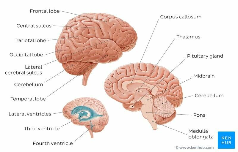
The central nervous system (CNS) consists of the brain and the spinal cord. The CNS controls most of the functions of the body including things like movement, planning and much more! The brain is the portion of the central nervous system that is contained within the skull. The cerebrum forms most of the brain and helps to perform the higher functions of the nervous system. It contains the frontal lobe, parietal lobe, temporal lobe and occipital lobe. The cerebellum is the part of the brain that moderates reflexes that coordinate voluntary movements
Brain Anatomy
The cerebrum is composed of the frontal lobe, the parietal lobe, the occipital lobe, and the temporal lobe. It is covered with gyri (ridges/folds) and sulci (depressions) in order to increase surface area. An enlarged cerebrum is unique to mammals and supports high level cognitive functioning.
The cerebellum regulates motor movements. It coordinates voluntary movements such as posture, balance, coordination, and speech. This results in smooth and balanced muscular activity.
Brain Stem
The brain stem controls the flow of messages between the brain and the rest of the body. It also controls basic body functions like breathing, swallowing, heart rate, blood pressure, and consciousness.

kenhub

https://www.funbiology.com/brain-stem-definition-structure-and-function/

Brain
The brain is composed of the cerebrum, the cerebellum, and the brain stem. It is important for coordinating body movement and function.

Skull
The skull can be divided into three parts: the braincase, the facial bones, and the mandible. The braincase provides support and protection to the brain. The facial bones support the face, teeth, nasal, and oral cavities. The division between the braincase and the facial bones is at the levels of the orbits of the eyes. The mandible is also known as the lower jaw or the jawbone.

Canine Organs
The Respiratory System
To understand the pleural membranes, let's use a "hand in the balloon analogy": Imagine holding a weakly inflated balloon (= a pleural sac) in the open palm of your left hand (= the chest wall). Imagine pushing your closed right fist (= a lung) into your left palm in such a way that you compress the balloon to the point that your fist can rub against your palm, separated only by two thin layers of balloon rubber. Now swivel your fist back and forth in your cupped palm (to simulate the movement of the lung against the inner surface of the chest wall with each breath). As long as the balloon was inflated “moistly” with a good amount of mucoid saliva (yuck!), it’s the two layers of balloon rubber that will glide smoothly against each other, rather than your two hands. This protects the fist (lung) from abrasion against the palm (chest wall). This is the system your lungs use to allow free, frictionless movement of the lungs during inhalation and exhalation.

Lungs
Lungs consist of a right and left side. Different species vary in the numbers of lobes they have. The dog’s lungs are deeply lobated, meaning they have many lobes. There are four lobes on the right side, and two lobes on the left side. The right lung consists of the cranial lobe, the caudal lobe, the middle lobe, and the accessory lobe. The left lung consists of the cranial lobe and caudal lobe.
Can you think of a reason there are less lung lobes on the left side?
There are less lung lobes on the left side to make room for the heart!
The Cardiovascular System
Oxygen poor blood enters the heart from the body via the cranial and caudal vena cava. Blood first enters the right atrium, and is pumped through the right atrioventricular valve into the right ventricle. From the right ventricle, the blood is moved through the pulmonary valve into the pulmonary trunk to the lungs to become oxygenated. Once oxygenated, the blood returns via the pulmonary vein and moves into the left atrium and is pumped through the left atrioventricular valve into the left ventricle. From the left ventricle, the blood is pumped through the aortic valve into the aorta which carries the oxygenated blood to the body.

https://moosmosis.org/2020/07/09/circulatory-system-blood-flow-pathway-through-the-heart/
The Gastrointestinal System

Canine Oral Cavity
Teeth are important for breaking down food into smaller more digestible pieces. Dogs and cats are carnivores, so they have teeth adapted for this lifestyle.
Dogs have a total of 42 teeth! On their upper jaw (maxilla), adult dogs have 6 incisors, 2 canines, 6 premolars, and 4 molars. On their bottom jaw (mandible), adult dogs have 6 incisors, 2 canines, 8 premolars, and 6 molars. Incisors are specially adapted for nibbling and grooming. Premolars are adapted for cutting, while molars are adapted for grinding.
Image Credit: http://www.germanwatchdogs.com/wp-content/uploads/2015/02/CanineDentition.jpg
Small Animal GI
The dog's digestive system consists of the same components as a human’s digestive system but it is organized slightly differently. Food travels through the mouth into the esophagus and down into the stomach. In the stomach, food is broken down by proteins called enzymes. From the stomach, food moves into the small intestine.
Despite its name, the small intestine is the largest part of the digestive tract. It is divided into three sections: the duodenum, the jejunum, and the ileum. It works with other organs in the digestive system to further digest food and absorb nutrients.
From the small intestine, food moves into the large intestine, also known as the colon. The colon absorbs water and propels feces toward the rectum for elimination.

VCA animal hospital
Anal sacs (often mistakenly referred to as anal glands) are two small structures located between the internal and external sphincter muscles of dogs and cats.
Each sac is lined with both sebaceous and apocrine glands that produce an odorous brown liquid.
As a dog or cat defaecates and relaxes their anal sphincter muscles, it puts pressure on the sacs, leading to the expulsion of their contents over the faeces.
In some cases the secretions do not release and build up in the sacs. This is more common in small dog breeds and can cause extreme discomfort to the dog.

VCA animal hospital

Stomach
The inside of both a dog’s and cat's stomach is divided into three sections: the fundus, the body, and the pylorus. The stomach also has large, linear folds called rugae. Rugae allow the stomach to stretch to accommodate large meals and help grip and move food during digestion.
The dog and cat's stomachs are completely glandular, meaning that the entire inner surface of the stomach is covered in glands that produce acid and other secretions that aid in digestion.
The Renal System
The kidneys function to remove wastes and extra fluids from the body. They help maintain a healthy balance of water, salts, and minerals in your blood. They also make hormones that help control blood pressure, make red blood cells, and keep bones strong and healthy.
Compared to other animals, like cows, cat and dog kidneys look pretty similar. However, dog kidneys are slightly larger! Can you see this in the images on the right?
Canine Kidney

Feline Kidney


